

specialties
Echocardiography
Cardiac Consultations
Echocardiography
Cardiac Catheterization & Angioplasty
Nuclear Cardiac Stress Text
Pacemaker Interrogation
follow-up
Lipid Management
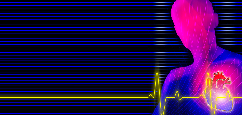
An echocardiogram is a sonogram of the heart, also known as cardiac ultrasound.
It uses standard ultrasound techniques to image two-dimensional slices of the heart. In addition to creating two-dimensional pictures of the cardiovascular system, an echocardiogram can also produce accurate assessment of the velocity of blood and cardiac tissue at any point. This allows assessment of cardiac valve areas and function, any abnormal communications between the left and right side of the heart, any leaking of blood through the valves (valvular regurgitation), and calculation of the cardiac output as well as the ejection fraction. Other parameters measured include cardiac dimensions and intracardiac pressures.
Echocardiography is used to diagnose cardiovascular diseases. In fact, it is one of the most widely used diagnostic tests for heart disease. It can provide a wealth of helpful information, including the size and shape of the heart, its pumping capacity and the location and extent of any damage to its tissues. It is especially useful for assessing diseases of the heart valves. It not only allows doctors to evaluate the heart valves, but it can detect abnormalities in the pattern of blood flow, such as the backward flow of blood through partly closed heart valves, known as regurgitation. Echocardiography is useful in the assessment of coronary artery disease. Echocardiography can also help detect hypertrophic cardiomyopathy. The biggest advantage to echocardiography is that it is noninvasive (doesn't involve breaking the skin or entering body cavities) and has no known risks or side effects.
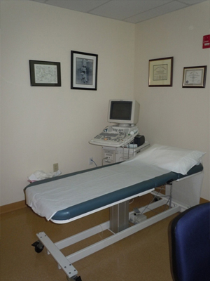
Transthoracic echocardiogram
A standard echocardiogram is also known as a transthoracic echocardiogram (TTE), or cardiac ultrasound. In this case, the echocardiography transducer (or probe) is placed on the chest wall of the subject, and images are taken through the chest wall. This is a non-invasive, highly accurate and quick assessment of the overall health of the heart. Transthoracic echocardiograms are performed at Princeton Interventional Cardiology. All Transthoracic echocardiograms are also read in-house within 24 hours by our board certified cardiologists.
Transesophageal Echocardiogram
This is an alternative way to perform an echocardiogram. A specialized probe containing an ultrasound transducer at its
tip is passed into the patient's esophagus. This allows image and Doppler evaluation which can be recorded. This is known
as a transesophageal echocardiogram or TEE. Transesophageal echocardiograms are performed at the University Medical
Center at Princeton.
Accreditation
United States: The "Intersocietal Commission for the Accreditation of Echocardiography Laboratories" (ICAEL) sets standards for echo labs, cardiologists and technologists in the US. Once all requirements have been met, the lab will receive ICAEL certification. Princeton Interventional Cardiology, P.A. maintains an ICAEL accreditation certificate for our echo lab since 2008. Dr. James R. Beattie is the Director of our Echocardiography Lab


Copyright 2011 Princeton Interventional Cardiology, P.A.: Cardiology Princeton NJ. All Rights Reserved.
389 Wall St Princeton, NJ 08540
(609) 921-2800