
specialties
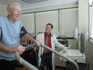
Nuclear Cardiac Stress Test
Cardiac Consultations
Echocardiography
Cardiac Catheterization & Angioplasty
Nuclear Cardiac Stress Text
Pacemaker Interrogation
follow-up
Lipid Management
Myocardial perfusion studies can thus identify areas of the heart muscle that have an inadequate blood supply as well as the areas of heart muscle that are scarred from a heart attack. In addition to the localization of the coronary artery with atherosclerosis, myocardial perfusion studies quantify the extent of the heart muscle with a limited blood flow and can also provide information about the pumping function of the heart. Thus, it is superior to routine exercise stress testing and provides the necessary information to help identify which patients are at an increased risk for a heart attack and may be candidates for invasive procedures such as coronary angiography, angioplasty and heart surgery.
Evaluation of Cardiac Function with Radionuclide Ventriculography
Radionuclide ventriculography is a noninvasive study, which provide information about the pumping function of the heart. In patients with coronary artery disease, and in those who have had a heart attack, the assessment of the pumping function of the heart (also known as the ejection fraction) is essential in the prediction of both long term and short-term survival. A small dose of an imaging agent is injected into the blood stream and pictures of the four chambers of the heart are taken using a special camera (gamma camera). These techniques can also provide information about the function of the valves of the heart, the integrity of all the cardiac chambers and can be used to monitor the effect of different drugs on the heart muscle (in patients with cancer who are treated with chemotherapy). The evaluation of cardiac function with radionuclide ventriculography is accurate and noninvasive and continues to play a critical role in predicting outcomes in patients with heart disease.
Nuclear cardiology studies continue to play an increasingly important role in the new millennium, in the noninvasive diagnosis of coronary artery disease, the assessment of the pumping function of the heart and in the prediction of outcomes in patients with heart disease.
Accreditation:
Today's health care organizations are held to high levels of accountability by patients, peers and by Medicare and other payers. The purpose of the Intersocietal Commission for the Accreditation of Nuclear Medicine Laboratories (ICANL) is to ensure high quality patient care and to promote health care by providing a mechanism to encourage and recognize the provision of quality nuclear cardiology and nuclear medicine by a voluntary accreditation. Laboratories attaining accreditation demonstrate a willingness to surpass expectations. Maintaining an ICANL accreditation demonstrates to our patients our commitment to providing the highest standard of care.
Princeton Interventional Cardiology, P.A. has maintained an ICANL accreditation of its nuclear cardiology laboratory since 2005. Dr. John Passalaris, a board, certified nuclear cardiologist is Director of the Nuclear Lab.
Nuclear Cardiology
Heart disease is the leading cause of death in the western world. Each year in the U.S.A, more than 500,000 men and women will die from coronary artery disease. During the past two decades, major strides have been made in the diagnosis and treatment of heart disease. Nuclear Cardiology has played a pivotal role in establishing the diagnosis of heart disease and in the assessment of disease extent and the prediction of outcomes in the setting of coronary artery disease.
Nuclear cardiology studies use noninvasive techniques to assess myocardial blood flow, evaluate the pumping function of the heart as well as visualize the size and location of a heart attack. Among the techniques of nuclear cardiology, myocardial perfusion imaging is the most widely used.
Myocardial Perfusion Imaging:
Myocardial perfusion images are combined with exercise to assess the blood flow to the heart muscle. Exercise is usually in the form of walking on the treadmill or riding a stationary bicycle. A "chemical" stress test using the drug dipyridamole can be performed in patients who are not able to exercise maximally, providing similar information about the heart's blood flow.
A small amount of an imaging agent, tetrofosmin (Myoview), is injected into the blood stream during rest and during exercise or chemical stress. A scanning device (gamma camera) is used to measure the uptake by the heart of the imaging material during exercise (or chemical stress) and at rest. If there is significant blockage of a coronary artery, the heart muscle may not get enough of a blood supply in the setting of exercise or during chemical stress. This decrease in blood flow will be detected by the camera.


Copyright 2011 Princeton Interventional Cardiology, P.A.: Cardiology Princeton NJ. All Rights Reserved.
389 Wall St Princeton, NJ 08540
(609) 921-2800